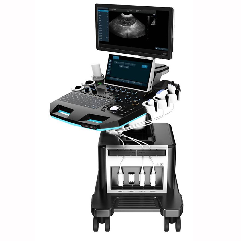
Trolley Veterinary Color Doppler Ultrasound Scanner VC-U9
VC-U9 Trolley Veterinary Color Doppler Ultrasound Scanner
- SKU
- VC-U9
- Brand
- MoreVET
- Manufactured by
- MoreVET
- Availability
- In stock
- ✓ Worldwide shipping


Clinical overview
Product overview
- Application
It’s applicable for animals’ examination and diagnosis on digestive system, reproductive system, urinary system for animal hospitals and scientific institutions.
- Technical Specifications
|
PARAMETER |
SPECIFICATION |
|
System technical specifications and summary |
|
|
Structure Style |
Dual Screen Cart |
|
Operating System |
Windows 10 |
|
Pulsed Wave Doppler Imaging (PW) |
|
|
Directional Power Doppler Imaging (DPDI) |
|
|
Real-time Triplex |
|
|
Spatial Compound Imaging |
|
|
Tissue Harmonic Imaging (THI) |
|
|
2B/4B Imaging Modes |
|
|
Support Language |
, English, French, Russian, Spanish |
|
Monitor sizes |
≥21.5 Inch |
|
All-in-one clipboard |
saved images display on the right of the screen, which can be directly transferred or deleted |
|
The system has the function of on-the-spot upgrade |
|
|
Preset table Conditions |
Preset optimal image inspection conditions to reduce adjustment during operation |
|
Support real-time 3D imaging |
|
|
Probe interface |
≥ 4 |
|
Trapezoidal Imaging |
|
|
One Key Smart Optimization |
|
|
Probe |
|
|
Convex probe |
Fundamental Frequency 2.5MHz/3.0MHz/3.5MHz/4.0MHz/H4.0MHz/H5.0MHz, six-segment frequency conversion (detecting depth: 30-255mm) |
|
Linear probe |
Fundamental Frequency 6.0MHz/7.5MHz/8.5MHz/10.0MHz/12.0MHz/H10.0MHz, six-segment frequency conversion (detecting depth 20-128mm) |
|
Phased probe |
Fundamental frequency 2.5MHz/3.0MHz/3.5MHz/4.0MHz/H3.0MHz/H4.0MHz.six-segment frequency conversion (detecting depth 100-244mm) |
|
Volume probe |
Fundamental frequency 2.0MHz/3.0MHz/4.5MHz/6.0MHz/H5.0MHz,five-segment frequency conversion (detecting depth 30-237mm) |
|
Micro-convex probe (R11) |
Fundamental frequency4.5MHz/6.0MHz/7.0MHz/9.0MHz/H8.0Mhz,five-segment frequency conversion (detecting depth 30-111mm) |
|
Rectal probe |
fundamental frequency 4.0MHz/6.5MHz/9.0MHz/H8.0MHz four-segment frequency conversion (detecting depth 20-110mm) |
|
High Frequency Phased Array |
Fundamental frequency 3.0MHz/5.0MHz/7.0MHz/H6.0MHz/H7.0MHz five-segment conversion (detecting depth 40-238mm) |
|
L25High Frequency linear probe |
Fundamental frequency 6.0MHz/7.5MHz/8.5MHz/10MHz/12MHz/H10.0MHz six-segment conversion (detecting depth 20-110mm) |
|
Backfat probe |
Fundamental frequency 2.0MHz/3.5MHz/4.0MHz/5.0MHz/H4.0MHz/H5.0MHz six-segment conversion (detecting depth 30-237mm) |
|
2D Imaging Mode |
|
|
Gain |
0-100 |
|
TGC |
8 segment adjustable |
|
Dynamic |
20-280, 20 levels adjustable |
|
Pseudo color |
0-11 level,visually adjustable |
|
Sound power |
Sound power: 5%-100%, step 5% visually adjustable |
|
Body mark |
≥18 kinds optional |
|
Maximum focus |
≥6, which can be moved throughout the whole process |
|
Grey scale map |
0-7, 7 levels adjustable |
|
Filter |
0-4 |
|
Scanning range |
50%-100% |
|
Frame correlation |
0-4, 4 level adjustable |
|
The screen has real-time display of sound power, probe frequency, dynamic range, pseudo color, gray scale and other 14 parameters can be adjusted |
|
|
Line density |
low, middle, high, 3 levels adjustable |
|
Noise reduction |
0-14 |
|
Noise reduction: 0-14 |
|
|
Color Frame correlation: 0-12, 12 levels visually adjustable |
|
|
Color map: 0-7, 7 levels visually adjustable |
|
|
Color flip: adjustable |
|
|
B C real-time split screen mode |
|
|
Base line: 11 levels, visually adjustable |
|
|
Line density: low, high, 2 levels adjustable |
|
|
Filter: 0-5 levels adjustable |
|
|
Spectral Doppler Imaging Mode |
|
|
Sampling volume angle correction |
-80°~80°adjustable |
|
Sample volume |
0.5mm-20mm adjustable |
|
Frequency |
2.5MHz, 3.0MHz |
|
Base line |
9 levels, adjustable |
|
Pseudo color |
0-5 |
|
Parameter display |
≥4 kinds, adjustable |
|
Speed scale |
7.2-231cm/s (different probes have different ranges) |
|
Spectrum envelope function |
real time automatic spectrum envelope, manual spectrum envelope, other. The system automatically analyses and displays various data such as PS, ED, RI, PI, S/D, HR, etc. |
|
Grey map |
0-7 |
|
Filter |
0-8 |
|
Dynamic range |
10-95db, step 5 |
|
Noise reduction |
0-28 |
|
Sound volume |
0-1007 |
|
3D imaging mode (optional) |
|
|
Quick Angle |
Support 0°, 90°, 180°, 270° rotation of the 3D window image |
|
Display layout |
Support “double””quad”, “single” image display |
|
Reconstruction mode |
Real-Skin, surface, Max, Min, XRax five reconstruction modes |
|
Pseudo-color display |
support 0-7 level adjustment |
|
Image magnification |
support level 5 |
|
Contrast |
0%-100% |
|
Threshold |
0%-100% |
|
Smooth |
≥3 levels adjustable |
|
X-axis, Y-axis, Z-axis rotation support adjustable |
|
|
Brightness |
0%-100% |
|
4D imaging mode (optional) |
|
|
Quick Angle |
Support 0°, 90°, 180°, 270° rotation of the 3D window image |
|
Display layout |
Support “double”, “quad”, “single” image display |
|
Reconstruction mode |
Real-Skin, surface, Max, Min, XRax five reconstruction modes |
|
Pseudo-color display |
support 0-7 level adjustment |
|
Image magnification |
support level 5 |
|
Contrast |
0%-100% |
|
Threshold |
0%-100% |
|
Smooth |
≥3 levels adjustable |
|
Pseudo-color |
≥7 levels adjustable |
|
X-axis, Y-axis, Z-axis rotation support adjustable |
|
|
Linear density |
support two-level adjustment |
|
Measurement and Analysis |
|
|
Measurement items include distance, area, angle, time, slope, heart rate, speed, acceleration, blood flow path, blood flow spectrum trace, resistance index/pulsatility index and other professional measurements |
|
|
Obstetrical measurement packages include dogs, cats, horses, cattle, sheep |
|
|
Measurement line color and line type can be adjusted freely (including active color and finished color) |
|
|
The display position and font size of the measurement results can be adjusted as needed. |
|
|
Professional software package |
abdomen, obstetrics, urology, etc. |
|
Image and text management system: image saving format: BMP DCM JPG |
|
|
Host built-in ≥ 256G solid state hard disk starts quickly and stably |
|
|
Movie playback |
≥ 600 frames |
|
Built-in file information management system |
can record the number, name, inspection number, inspection date, etc., can search and manage the number, inspection number, name, etc |
|
Report types ≥ 6. Provide photo proof. |
|
|
One-click quick report image and text management |
|
|
Interface |
|
|
USB ports, 1 Video, 1 S-Video, 1 DVI, 1 HDMI, 1 RJ-45. |
|
|
Configuration |
|
|
Color Doppler Ultrasonic Diagnosis System Host 1 |
|
|
Probe |
micro-convex R11 (optional), convex array probe (optional), linear array probe (optional), micro-convex R15 probe (optional), cardiac probe (optional), cavity probe (optional) volume probe (optional), etc. |
|
Video printer (optional), laser printer (optional), etc. |
|
Technical specifications
Engineering at a glance
Documentation, IFU and DOCs available on request — contact our sales team for the full regulatory file.
- Structure Style
- Dual Screen Cart
- Operating System
- Windows 10
- Support Language
- English, French, Russian, Spanish
- Monitor sizes
- ≥21.5 Inch
- All-in-one clipboard
- saved images display on the right of the screen, which can be directly transferred or deleted
- Preset table Conditions
- Preset optimal image inspection conditions to reduce adjustment during operation
- Probe interface
- ≥ 4
- Convex probe
- Fundamental Frequency 2.5MHz/3.0MHz/3.5MHz/4.0MHz/H4.0MHz/H5.0MHz, six-segment frequency conversion (detecting depth: 30-255mm)
- Linear probe
- Fundamental Frequency 6.0MHz/7.5MHz/8.5MHz/10.0MHz/12.0MHz/H10.0MHz, six-segment frequency conversion (detecting depth 20-128mm)
- Phased probe
- Fundamental frequency 2.5MHz/3.0MHz/3.5MHz/4.0MHz/H3.0MHz/H4.0MHz.six-segment frequency conversion (detecting depth 100-244mm)
- Volume probe
- Fundamental frequency 2.0MHz/3.0MHz/4.5MHz/6.0MHz/H5.0MHz,five-segment frequency conversion (detecting depth 30-237mm)
- Micro-convex probe (R11)
- Fundamental frequency4.5MHz/6.0MHz/7.0MHz/9.0MHz/H8.0Mhz,five-segment frequency conversion (detecting depth 30-111mm)
- Rectal probe
- fundamental frequency 4.0MHz/6.5MHz/9.0MHz/H8.0MHz four-segment frequency conversion (detecting depth 20-110mm)
- High Frequency Phased Array
- Fundamental frequency 3.0MHz/5.0MHz/7.0MHz/H6.0MHz/H7.0MHz five-segment conversion (detecting depth 40-238mm)
- L25High Frequency linear probe
- Fundamental frequency 6.0MHz/7.5MHz/8.5MHz/10MHz/12MHz/H10.0MHz six-segment conversion (detecting depth 20-110mm)
- Backfat probe
- Fundamental frequency 2.0MHz/3.5MHz/4.0MHz/5.0MHz/H4.0MHz/H5.0MHz six-segment conversion (detecting depth 30-237mm)
- Gain
- 0-100
- TGC
- 8 segment adjustable
- Dynamic
- 20-280, 20 levels adjustable
- Pseudo color
- 0-11 level,visually adjustable
- Sound power
- Sound power: 5%-100%, step 5% visually adjustable
- Body mark
- ≥18 kinds optional
- Maximum focus
- ≥6, which can be moved throughout the whole process
- Grey scale map
- 0-7, 7 levels adjustable
- Filter
- 0-4
- Scanning range
- 50%-100%
- Frame correlation
- 0-4, 4 level adjustable
- Line density
- low, middle, high, 3 levels adjustable
- Noise reduction
- 0-14
- Sampling volume angle correction
- -80°~80°adjustable
- Sample volume
- 0.5mm-20mm adjustable
- Frequency
- 2.5MHz, 3.0MHz
- Base line
- 9 levels, adjustable
- Pseudo color
- 0-5
- Parameter display
- ≥4 kinds, adjustable
- Speed scale
- 7.2-231cm/s (different probes have different ranges)
- Spectrum envelope function
- real time automatic spectrum envelope, manual spectrum envelope, and other. The system automatically analyses and displays various data such as PS, ED, RI, PI, S/D, HR, etc.
- Grey map
- 0-7
- Filter
- 0-8
- Dynamic range
- 10-95db, step 5
- Noise reduction
- 0-28
- Sound volume
- 0-1007
- Quick Angle
- Support 0°, 90°, 180°, 270° rotation of the 3D window image
- Display layout
- Support “double””quad”, “single” image display
- Reconstruction mode
- Real-Skin, surface, Max, Min, XRax five reconstruction modes
- Pseudo-color display
- support 0-7 level adjustment
- Image magnification
- support level 5
- Contrast
- 0%-100%
- Threshold
- 0%-100%
- Smooth
- ≥3 levels adjustable
- Brightness
- 0%-100%
- Quick Angle
- Support 0°, 90°, 180°, 270° rotation of the 3D window image
- Display layout
- Support “double”, “quad”, “single” image display
- Reconstruction mode
- Real-Skin, surface, Max, Min, XRax five reconstruction modes
- Pseudo-color display
- support 0-7 level adjustment
- Image magnification
- support level 5
- Contrast
- 0%-100%
- Threshold
- 0%-100%
- Smooth
- ≥3 levels adjustable
- Pseudo-color
- ≥7 levels adjustable
- Linear density
- support two-level adjustment
- Professional software package
- abdomen, obstetrics, urology, etc.
- Movie playback
- ≥ 600 frames
- Built-in file information management system
- can record the number, name, inspection number, inspection date, etc., and can search and manage the number, inspection number, name, etc
- Probe
- micro-convex R11 (optional), convex array probe (optional), linear array probe (optional), micro-convex R15 probe (optional), cardiac probe (optional), cavity probe (optional) , volume probe (optional), etc.
Documents & downloads
Regulatory paperwork & manuals
Pre-cleared documentation for this device. Full tender-pack originals are available on request.
Documents & downloads
Regulatory paperwork & manuals
This device's IFU, CE Declaration of Conformity, ISO 13485 certificate and (where applicable) FDA 510(k) clearance ship with the order. Reach out below for the pack ahead of purchase.
Get in touch
Need configuration help, certificates, or volume pricing?
Our clinical sales team will scope quantities, voltage, accessories and regulatory paperwork — usually within one business day.
Customer reviews
No reviews yet — be the first to share your experience.
Related products
See all →
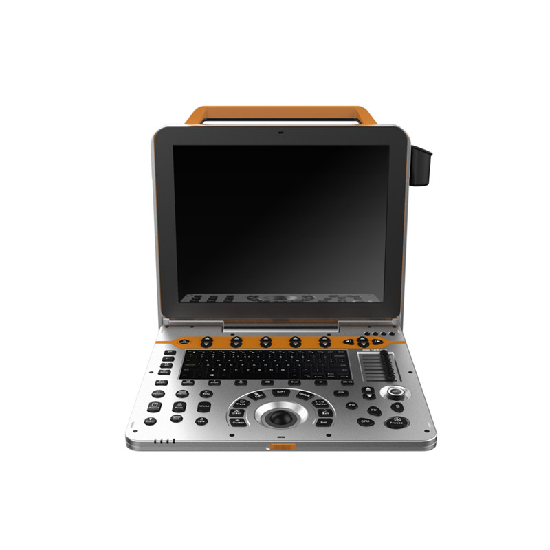
Portable Veterinary Color Doppler Ultrasound Scanner VC-E80
VC-E80 Portable Veterinary Color Doppler Ultrasound Scanner
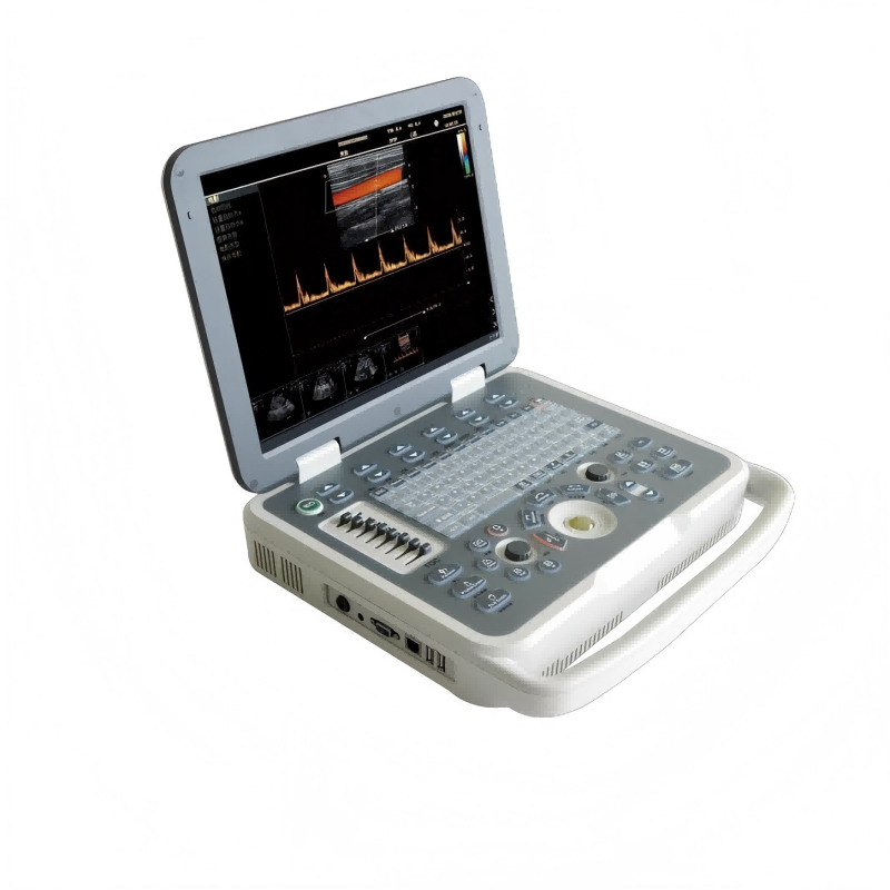
Portable Veterinary Color Doppler Ultrasound Scanner VC-U60
VC-U60 Portable Veterinary Color Doppler Ultrasound Scanner

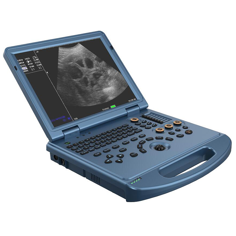
Portable Color Doppler Ultrasound Scanner VC-U6
VC-U6 Portable Color Doppler Ultrasound Scanner
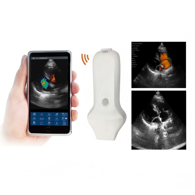
Phased Array Probe Type Wireless Color Doppler Scanner VC-4PA
VC-4PA Phased Array Probe Type Wireless Color Doppler Scanner
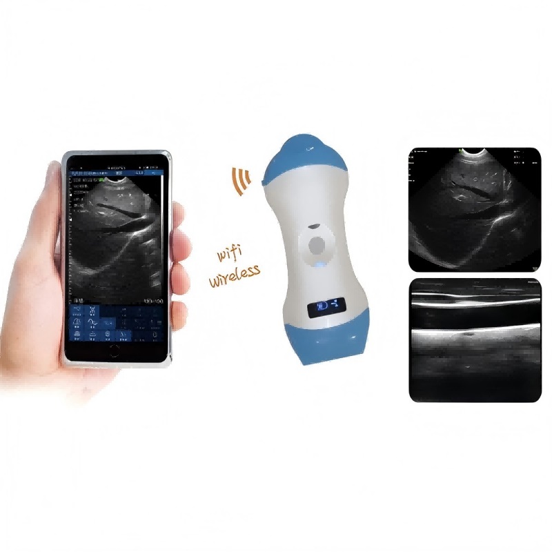
Micro-Convex+Linear Probe Type Wireless Color Doppler Scanner VC-3ML
VC-3ML Micro-convex+Linear Probe Type Wireless Color Doppler Scanner
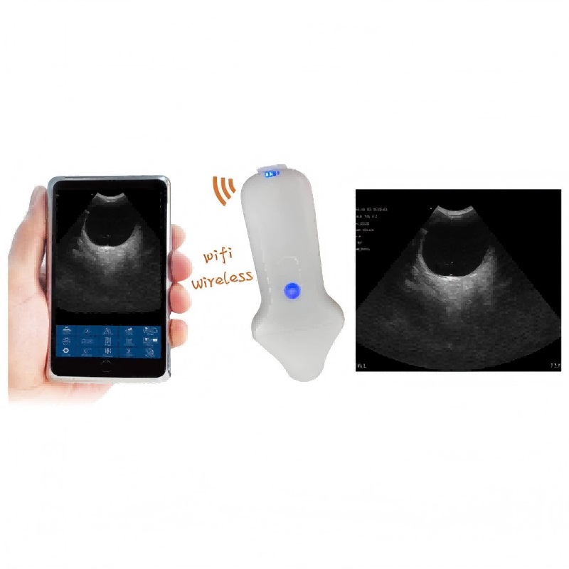
Micro-convex Probe Type Wireless Color Doppler Scanner VC-3M
VC-3M Micro-convex Probe Type Wireless Color Doppler Scanner
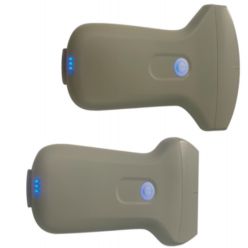
Convex/ Linear Probe Type Wireless Ultrasound Scanner VC-3CE/ VC-3LE
VC-3CE/ VC-3LE Convex/ Linear Probe Type Wireless Ultrasound Scanner