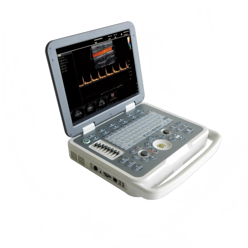
Portable Veterinary Color Doppler Ultrasound Scanner VC-E80
VC-E80 Portable Veterinary Color Doppler Ultrasound Scanner
- SKU
- VC-E80
- Brand
- MoreVET
- Manufactured by
- MoreVET
- Availability
- In stock
- ✓ Worldwide shipping


Clinical overview
Product overview
Features
-A new ultrasound diagnostic platform with Innovations in areas of digital electronics.
-Achieve a new level of ultrasound diagnostic precision and higher diagnostic confidence.
-A revolutionary workflow control is provided with the user-centric architecture of the new software platform.
Technical Specifications
|
PARAMETER |
SPECIFICATION |
|
Technical platform:linux +ARM+FPGA |
|
|
Monitor |
|
|
15-inch, high resolution, progressive scan, Wide Angle of view |
|
|
Resolution |
1024*768 pixels
|
|
Image display area |
640*480
|
|
Hard disk:Internal 128GB hard disk for patient database management Allow storage of patient studies that include images,clips,reports and measurements |
|
|
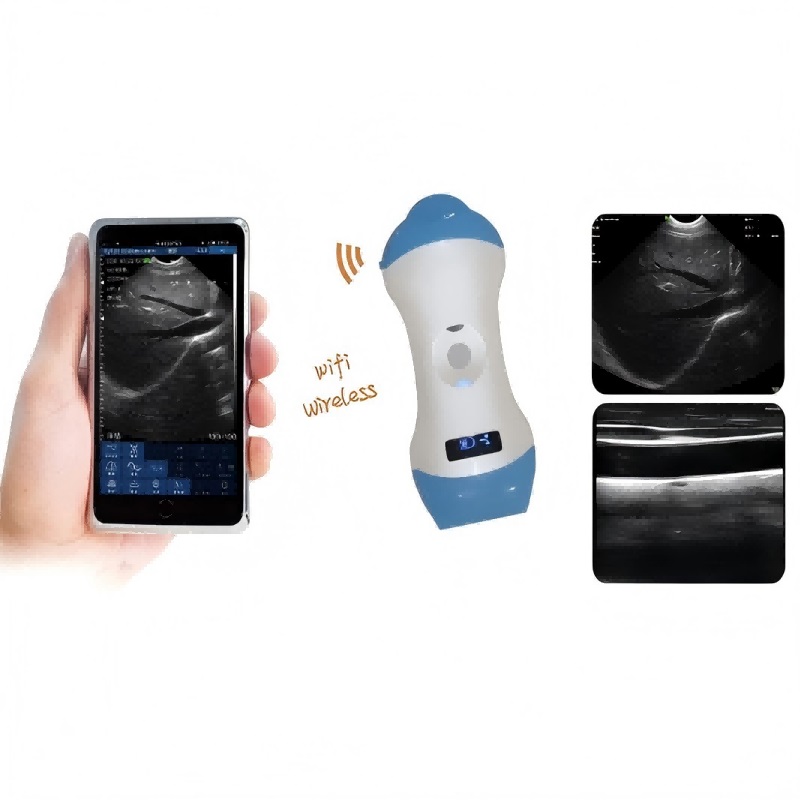
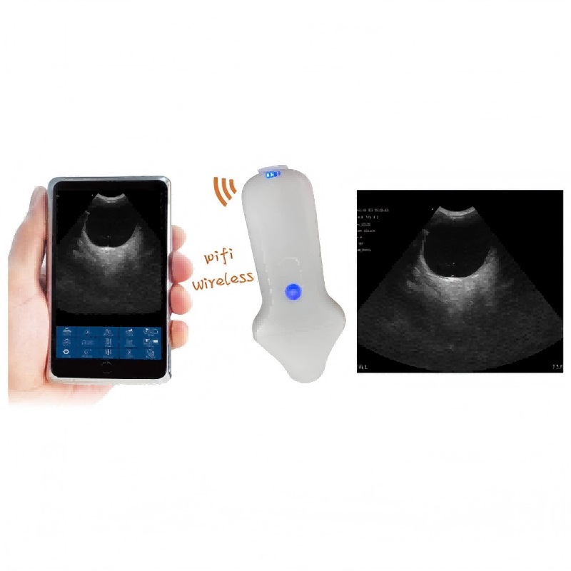
Transducer Ports |
Two active universal transducer ports that support standard(curved array, linear array)Probe,Unique industrial design provides easy access to all transducer ports |
|
Probe available |
|
|
3C6A |
3.5MHz/R60/80,Convex array probe |
|
7L4A |
7.5MHz/L38mm/80,Convex array probe |
|
6E1A |
6.5MHz/R10/80,EndocavityConvex array probe |
|
6C15A |
6.5MHz/R15/80,Micro convex array probe |
|
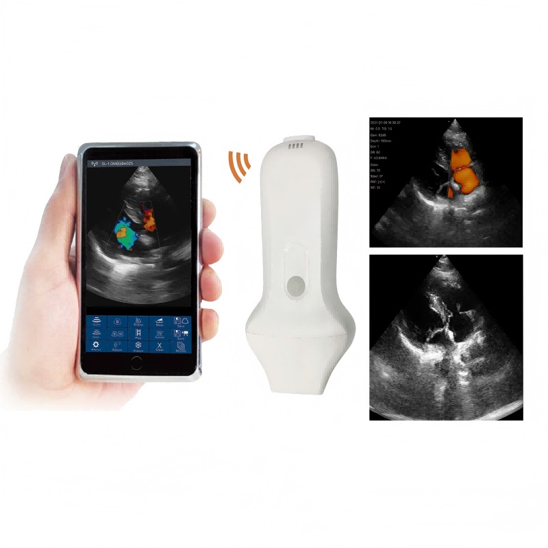
7 Imaging modes |
|
|
B-mode |
Fundamental and Tissue harmonic imaging Color Flow Mapping (Color) B/BC Dual Real-Time Power Doppler Imaging (PDI) PW Doppler M-mode |
|
frequency number B/M |
|
|
Fundamental wave |
≥3 |
|
harmonic wave |
≥2 |
|
Color/PDI |
≥2 |
|
PW |
≥2 |
|
Cine |
|
|
B mode |
≥5000frames |
|
B+Color/B+PDI mode |
≥2300frames |
|
M、PW |
≥ 190s |
|
image zoom |
available on live, 2B, 4B and reviewed images up to 10X zoom |
|
image save |
format BMP、JPG、FRM(single image); CIN、AVI(multiple images) Support DICOM, conform to DICOM3.0 standard Built in workstation,support patient data search and browse |
|
language
|
Support English、Spanish、French、German、Czech、Russian languages. Can be easily extended to support other languages |
|
Battery |
Built in large capacity lithium battery, working condition. Continuous working time ≥ 1 hours. Screen provides power display information |
|
Other functions |
Comment、BodyMark、Biopsy、PICC、★Lito、★IMT、★Report template、★Support,USB mouse, etc |
|
Imaging Parameters Bmode |
|
|
Dynamic range |
0-100% 5% step |
|
SpeckleReduction |
8 levels(0-7) |
|
ScanDensity |
H ,M ,L |
|
Gain |
0~100 % 2% step |
|
TGC |
eight TGC controls |
|
FrameAverage |
8 levels(0-7) |
|
LineAverage |
8 levels(0-7) |
|
Edge Enhance |
8 levels(0-7) |
|
Gray Maps |
15 types(0-14) |
|
Pseudocolor Maps |
7 types(0-6) |
|
Thermal Index |
TIC,TIS,TIB |
|
2B, 4B formats |
/ |
|
Invert (U/ D) and transposed (L/ R) |
/ |
|
Focus Number |
4 |
|
Focus Depth |
16 levels(depth and probe dependent) |
|
FOV |
5 levels |
|
Image depth up to 35 cm in 0.5~4cm increments (depth dependent) |
|
|
Phase inversion harmonic imaging technique is available for all probes |
|
|
Color mode |
|
|
Frequency |
2 levels |
|
Gain |
0~100% 2% steps |
|
Wall filter |
8 levels(0-7) |
|
Sensitivity |
H, M, L |
|
Flow |
H, M, L |
|
Packet Size1 |
5 levels(0-4) |
|
FrameAverage |
8 levels(0-7) |
|
PostProc |
4 levels(0-3) |
|
Invert |
On/Off |
|
Baseline |
7 levels(0-6) |
|
Color Maps |
4 levels(0-3) |
|
Color/PDI Width |
10%-100%, 10% |
|
Color/PDI Height |
0.5-30cm(probe dependent) |
|
Color/PDI Center Depth |
1-16cm(probe dependent) |
|
Steer |
+/-12 。,7 。(linear probe) |
|
PDI mode |
|
|
Frequency |
2 levels |
|
Gain |
0~100% 2% steps |
|
Wall filter |
8 levels(0-7) |
|
Sensitivity |
H, M, L |
|
Flow |
H, M, L |
|
Packet Size1 |
5 levels(0-4) |
|
FrameAverage |
8 levels(0-7) |
|
PostProc |
4 levels(0-3) |
|
Invert |
On/Off |
|
Baseline |
7 levels(0-6) |
|
PDI Maps |
2 levels(0-1) |
|
Color/PDI Width |
10%-100%, 10% |
|
Color/PDI Height |
0.5-30cm(probe dependent) |
|
Color/PDI Center Depth |
1-16cm(probe dependent) |
|
Steer |
+/-12 °, +/-7 °(linear probe) |
|
PW mode |
|
|
Frequency |
2 levels |
|
Sweep speed |
5 levels(0-4) |
|
Scale |
16 levels(0-15)(depth and probe dependent) |
|
Scale Unit |
cm/s,KHz |
|
Smooth |
8 levels(0-7) |
|
Pseudocolor Maps |
7 types(0-6) |
|
Dynamic range |
24-100, 2 step |
|
Gain |
0-100%, 2% step |
|
Wall filter |
4 levels(0-3) |
|
Dynamic range |
24-100, 2 step |
|
Gain |
0-100%, 2% step |
|
Wall filter |
4 levels(0-3) |
|
Angle correction |
-89 °+89 °, 1 °step |
|
Gate size |
8 levels(0-7mm) |
|
Wall filter |
5 levels(0-4) |
|
Invert |
On/Off |
|
Baseline |
7 levels |
|
Real-time auto Doppler trace: maximum velocity, mean velocity |
|
|
MMode |
|
|
Frequency |
Up to 3 fundamental and 2 harmonic imaging frequencies |
|
Edge enhance |
8 levels(0-7) |
|
Dynamic range |
0-100%,step 5% |
|
Gain |
0-100 ,step 2 |
|
Gray Maps |
15 levels(0-14) |
|
Pseudocolor Maps |
7 (0-6) |
|
Sweep speed |
5 levels(0-4) |
|
Image parameter save and restore user can press one key to save image parameters in screen user can press one key to restore image parameters to default status.
|
|
|
Ergonomic Design Frequently used controls center around the trackball Control panel is back lighted, waterproof and antisepticised Two USB ports are at the back of the system, which is more convenient for use
|
|
|
Exam Modes |
Abdomen Obstetrics Gynecology Fetal Heart Small parts Urology Carotid Thyroid Breast Vascular Kidney Pediatrics |
|
Product configuration |
|
|
Standard configuration |
Host( Built-in 128G hard disk) Adapter User’s Manual Power cable
|
|
Optional Accessories |
|
|
7E 10 Endocavity Convex probe |
|
|
3C25 Micro convex probe |
|
|
Convex probe |
|
|
Rectal probe |
|
|
Linear Probe |
|
|
USB report printer |
|
|
B/W or color Video printer |
|
|
Puncture rack |
|
|
Trolley |
|
Technical specifications
Engineering at a glance
Documentation, IFU and DOCs available on request — contact our sales team for the full regulatory file.
- Resolution
- 1024*768 pixels
- Image display area
- 640*480
- Transducer Ports
- Two active universal transducer ports that support standard(curved array, linear array)Probe,Unique industrial design provides easy access to all transducer ports
- 3C6A
- 3.5MHz/R60/80,Convex array probe
- 7L4A
- 7.5MHz/L38mm/80,Convex array probe
- 6E1A
- 6.5MHz/R10/80,EndocavityConvex array probe
- 6C15A
- 6.5MHz/R15/80,Micro convex array probe
- B-mode
- Fundamental and Tissue harmonic imaging Color Flow Mapping (Color) B/BC Dual Real-Time Power Doppler Imaging (PDI) PW Doppler M-mode
- Fundamental wave
- ≥3
- harmonic wave
- ≥2
- Color/PDI
- ≥2
- PW
- ≥2
- B mode
- ≥5000frames
- B+Color/B+PDI mode
- ≥2300frames
- M、PW
- ≥ 190s
- image zoom
- available on live, 2B, 4B and reviewed images up to 10X zoom
- image save
- format BMP、JPG、FRM(single image); CIN、AVI(multiple images) Support DICOM, conform to DICOM3.0 standard Built in workstation,support patient data search and browse
- language
- Support English、Spanish、French、German、Czech、Russian languages. Can be easily extended to support other languages
- Battery
- Built in large capacity lithium battery, working condition. Continuous working time ≥ 1 hours. Screen provides power display information
- Other functions
- Comment、BodyMark、Biopsy、PICC、★Lito、★IMT、★Report template、★Support,USB mouse, etc
- Dynamic range
- 0-100% ,5% step
- SpeckleReduction
- 8 levels(0-7)
- ScanDensity
- H ,M ,L
- Gain
- 0~100 % ,2% step
- TGC
- eight TGC controls
- FrameAverage
- 8 levels(0-7)
- LineAverage
- 8 levels(0-7)
- Edge Enhance
- 8 levels(0-7)
- Gray Maps
- 15 types(0-14)
- Pseudocolor Maps
- 7 types(0-6)
- Thermal Index
- TIC,TIS,TIB
- 2B, 4B formats
- /
- Invert (U/ D) and transposed (L/ R)
- /
- Focus Number
- 4
- Focus Depth
- 16 levels(depth and probe dependent)
- FOV
- 5 levels
- Frequency
- 2 levels
- Gain
- 0~100% ,2% steps
- Wall filter
- 8 levels(0-7)
- Sensitivity
- H, M, L
- Flow
- H, M, L
- Packet Size1
- 5 levels(0-4)
- FrameAverage
- 8 levels(0-7)
- PostProc
- 4 levels(0-3)
- Invert
- On/Off
- Baseline
- 7 levels(0-6)
- Color Maps
- 4 levels(0-3)
- Color/PDI Width
- 10%-100%, 10%
- Color/PDI Height
- 0.5-30cm(probe dependent)
- Color/PDI Center Depth
- 1-16cm(probe dependent)
- Steer
- +/-12 。,7 。(linear probe)
- Frequency
- 2 levels
- Gain
- 0~100% ,2% steps
- Wall filter
- 8 levels(0-7)
- Sensitivity
- H, M, L
- Flow
- H, M, L
- Packet Size1
- 5 levels(0-4)
- FrameAverage
- 8 levels(0-7)
- PostProc
- 4 levels(0-3)
- Invert
- On/Off
- Baseline
- 7 levels(0-6)
- PDI Maps
- 2 levels(0-1)
- Color/PDI Width
- 10%-100%, 10%
- Color/PDI Height
- 0.5-30cm(probe dependent)
- Color/PDI Center Depth
- 1-16cm(probe dependent)
- Steer
- +/-12 °, +/-7 °(linear probe)
- Frequency
- 2 levels
- Sweep speed
- 5 levels(0-4)
- Scale
- 16 levels(0-15)(depth and probe dependent)
- Scale Unit
- cm/s,KHz
- Smooth
- 8 levels(0-7)
- Pseudocolor Maps
- 7 types(0-6)
- Dynamic range
- 24-100, 2 step
- Gain
- 0-100%, 2% step
- Wall filter
- 4 levels(0-3)
- Dynamic range
- 24-100, 2 step
- Gain
- 0-100%, 2% step
- Wall filter
- 4 levels(0-3)
- Angle correction
- -89 °+89 °, 1 °step
- Gate size
- 8 levels(0-7mm)
- Wall filter
- 5 levels(0-4)
- Invert
- On/Off
- Baseline
- 7 levels
- Frequency
- Up to 3 fundamental and 2 harmonic imaging frequencies
- Edge enhance
- 8 levels(0-7)
- Dynamic range
- 0-100%,step 5%
- Gain
- 0-100 ,step 2
- Gray Maps
- 15 levels(0-14)
- Pseudocolor Maps
- 7 (0-6)
- Sweep speed
- 5 levels(0-4)
- Exam Modes
- Abdomen Obstetrics Gynecology Fetal Heart Small parts Urology Carotid Thyroid Breast Vascular Kidney Pediatrics
- Standard configuration
- Host( Built-in 128G hard disk) Adapter User’s Manual Power cable
Documents & downloads
Regulatory paperwork & manuals
Pre-cleared documentation for this device. Full tender-pack originals are available on request.
Documents & downloads
Regulatory paperwork & manuals
This device's IFU, CE Declaration of Conformity, ISO 13485 certificate and (where applicable) FDA 510(k) clearance ship with the order. Reach out below for the pack ahead of purchase.
Get in touch
Need configuration help, certificates, or volume pricing?
Our clinical sales team will scope quantities, voltage, accessories and regulatory paperwork — usually within one business day.
Customer reviews
No reviews yet — be the first to share your experience.
Related products
See all →
Trolley Veterinary Color Doppler Ultrasound Scanner VC-U9
VC-U9 Trolley Veterinary Color Doppler Ultrasound Scanner
Portable Veterinary Color Doppler Ultrasound Scanner VC-U60
VC-U60 Portable Veterinary Color Doppler Ultrasound Scanner

Portable Color Doppler Ultrasound Scanner VC-U6
VC-U6 Portable Color Doppler Ultrasound Scanner
Phased Array Probe Type Wireless Color Doppler Scanner VC-4PA
VC-4PA Phased Array Probe Type Wireless Color Doppler Scanner
Micro-Convex+Linear Probe Type Wireless Color Doppler Scanner VC-3ML
VC-3ML Micro-convex+Linear Probe Type Wireless Color Doppler Scanner
Micro-convex Probe Type Wireless Color Doppler Scanner VC-3M
VC-3M Micro-convex Probe Type Wireless Color Doppler Scanner
Convex/ Linear Probe Type Wireless Ultrasound Scanner VC-3CE/ VC-3LE
VC-3CE/ VC-3LE Convex/ Linear Probe Type Wireless Ultrasound Scanner