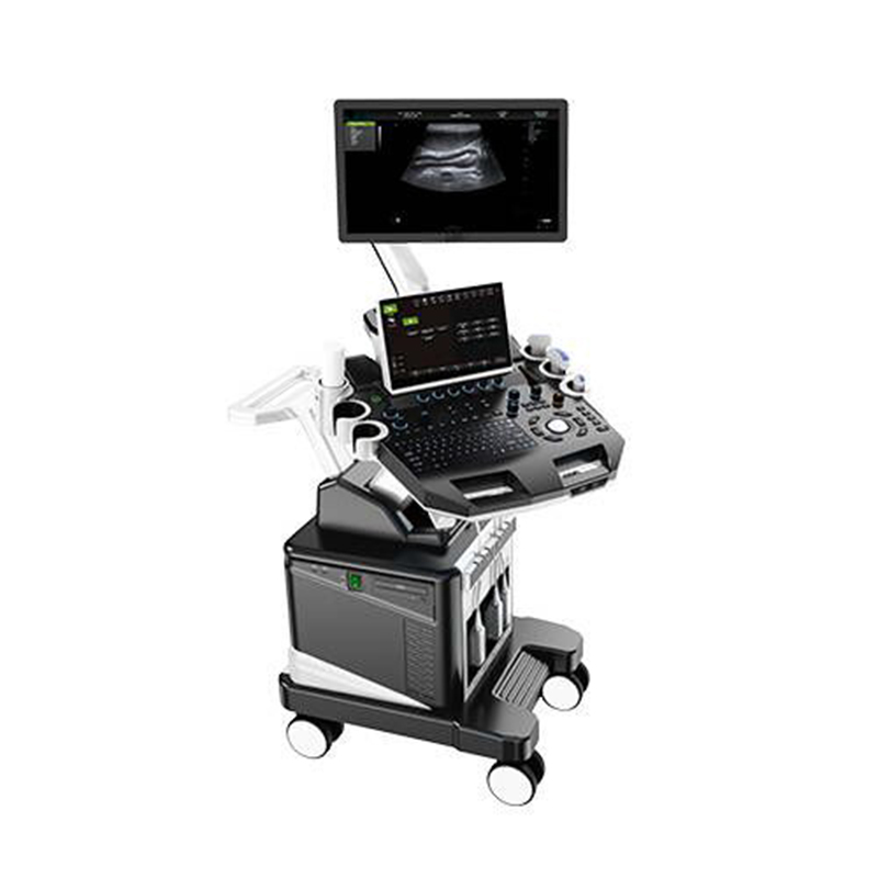
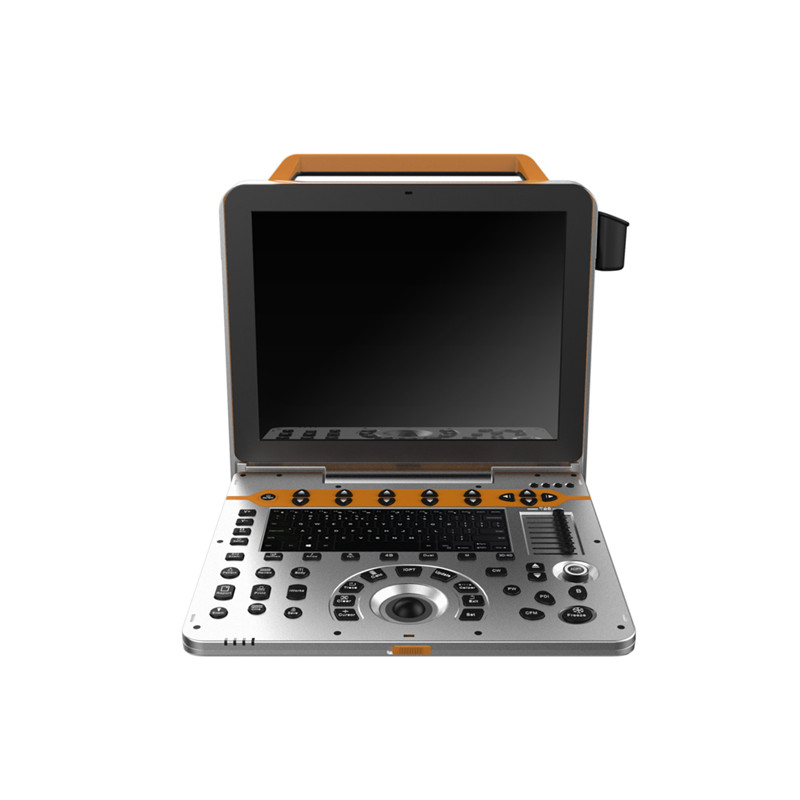
Echocardiography Trolley Color Doppler Ultrasound Scanner VC-U18
VC-U18 Echocardiography Trolley Color Doppler Ultrasound Scanner
- SKU
- VC-U18
- Brand
- MoreVET
- Manufactured by
- MoreVET
- Availability
- In stock
- ✓ Worldwide shipping


Clinical overview
Product overview
Features
- Ultrasonic host operating system: Windows operating system.
- carotid artery intima measurement thickness (IMT).
- Automatic spectral envelope measurement.
- Monitor: ≥5 inch, high definition ultrasonic display, ≥13.3 inch touch screen.
- Applications: Abdomen, obstetrics, gynecology, heart, urinary system, small organs, superficial, blood vessels, pediatrics, newborns, musculoskeletal.
- Applications and report: Abdominal,OB,GYN,Cardiac,Urinary,Small Parts,Superficial, Vascular, Pediatrics, Advanced measurement software packages, report software packages, case management software packages, etc
Technical Specifications
|
PARAMETER |
SPECIFICATION |
|
2D Imaging Mode |
|
|
Gain |
0-100, Step 1 adjustable |
|
TGC |
8 segment adjustable |
|
Dynamic |
30-180, 35 level, step 5 adjustable |
|
Speckle reduction |
0-5, 5 level |
|
Space Synthesis |
0-2, 2 level (Liner probe: 3 level, cardiac probe:0) |
|
Sound power |
2-10, 9 level |
|
Edge Enhancement |
0-5, 5 level |
|
Maximum focus point |
≥7, which can be moved throughout the whole process |
|
Grey scale |
0-67, 67 level |
|
False color |
0-67, 67 level |
|
Image style |
Soft-Comparison, 2 level |
|
Frame correlation |
0-4, 4 level adjustable |
|
Line density |
low, middle, high, 3 level |
|
Noise reduction |
0-5, 5 level |
|
The screen has real-time display of voice power, probe frequency, dynamic range, pseudo color, gray scale and other 11 parameters can be adjusted. |
|
|
Color Doppler Imaging Mode |
|
|
Blood gain |
0-100, Step 2 |
|
Parameter display |
Velocity, Variance |
|
B-Restrain (B/W restrain) |
0-7, 7 level |
|
Speed Through |
0-8, 8 level |
|
Sampling number |
6-24, 7 level |
|
Blood flow preferred |
0-8, 8 level |
|
Filtering |
1-6, 6 level |
|
Sound power |
2-6, 4 level |
|
Noise reduction |
0-4, 4 level |
|
Smooth treatment |
0-4, 4 level |
|
Frame correlation |
0-6, 6 level |
|
Chromatography (Blood flow graph) |
0-37, 37 level |
|
Line density |
Low-Middle-High, 3 level |
|
Frequency |
4 level adjustable |
|
Velocity |
Minimum 0.4K, Maximum 40.5K |
|
Convex probe |
0.4K-4.3K-38.5K |
|
Linear probe |
0.4K-14.7K-39.0K |
|
Trans-vaginal probe |
0.4K-7.8K-39.7K |
|
Volume probe |
0.4K-4.2K-34.8K |
|
Micro-convex probe |
0.4K-10.3K-40.5K |
|
Cardiac probe |
0.4K-7.8K-39.7K |
|
PS: The frequency of the probe changes and the frequency value changes |
|
|
PS: Frame rate changes with speed |
|
|
Pulse wave Doppler (PW) |
|
|
Gain |
0-100, Step 2 |
|
Spectrum envelope function: real time automatic spectrum envelope, manual spectrum envelope, and other modes. The system automatically analyses and displays various data such as PSV, EDV, RI, PI, S/D, ACC, HR and so on. Can wake up or close. |
|
|
Sample volume |
0.5mm- 30mm |
|
Blood angel |
-75—75 degree, Step 5 |
|
False color |
0-67, 67 level |
|
Dynamic range |
20-40, 4 level |
|
Filter |
0-9, 9 level |
|
Smooth treatment |
1-4, 4 level |
|
Sound power |
2-5, 4 level |
|
Volume |
0-100, 10 level, Step 10 |
|
Audio filtering |
0-4, 4 level |
|
Base line |
-1.0~1.0 |
|
Grey map |
0-67, 67 level |
|
Scan velocity |
100-500, 6 level |
|
PRF |
Minimum 0.5K, Maximum 87.5K |
|
Convex probe |
0.5K-4.3K-63.3K |
|
Linear probe |
0.5K-14.5K-78.4K |
|
Trans-vaginal probe |
0.5K-8.1K-78.4K |
|
Volume probe |
0.5K-4.2K-53.8K |
|
Micro-convex probe |
0.5K-10.3K-81.1K |
|
Cardiac probe |
0.5K-4.3K-87.5K |
|
Frequency |
4 level |
|
PS: The frequency of the probe changes and the PRF value changes |
|
|
PS: The frequency of the probe changes and the frequency value changes |
|
|
Continuous Wave Doppler (CW) |
|
|
Support probe |
Cardiac probe |
|
Adjustment of B mode parameters is switchable |
|
|
Gain |
0-100, Step 2 |
|
Sampling line position is adjustable |
|
|
PRF |
0.9K~36.1K |
|
Baseline |
-1.0~1.0 |
|
Blood angel |
-75~75 degree |
|
Grey map |
0-67 |
|
Scan velocity |
100-300 |
|
False color |
0-67 |
|
Dynamic range |
20-40 |
|
Filtering |
0-9, 9 level |
|
Smooth treatment |
1-4 |
|
Frequency |
2.0MHz/2.3MHz/2.5MHz/3.0MHz, 4 level adjustable |
|
Sound power |
2-5 |
|
Volume |
0-100 |
|
Audio Filtering |
0-4 |
|
Anatomical M imaging |
|
|
Support probe |
Convex probe, Linear probe,Cardiac probe |
|
Adjustment of B mode parameters is switchable |
|
|
Gain |
0-100, Step 2 |
|
M Sampling line angel is adjustable |
|
|
M Sampling line length is adjustable |
|
|
Sampling line: 3, Can be displayed or hidden separately |
|
|
Blood flow M mode (MC) |
|
|
Adjustment of B mode parameters is switchable |
|
|
Gain |
0-100, Step2 |
|
MC Sampling line angel is adjustable |
|
|
MC Sampling line length is adjustable |
|
|
Frequency |
4 level |
|
Sampling number |
6-24 |
|
Speed through |
0-8, 8 level |
|
Scan velocity |
150-500 |
|
Frame correlation |
0-6, 6 level |
|
Filtering |
1-6, 6 level |
|
Blood flow preferred |
0-8, 8 level |
|
Smooth treatment |
0-4, 4 level |
|
Map |
0-37, 37 level |
|
Elastography |
|
|
Adjustment of B mode parameters is switchable |
|
|
Gain |
0-100, Step 2 |
|
B/E, Double real-time display on the same screen |
|
|
Probe displacement curve display |
Up/Down |
|
Pressure indicator bar display |
|
|
Frequency |
8-9 level, Adjustable; According to the probe display |
|
Noise reduction |
0-2, 2 level |
|
Frame correlation |
0-3, 3 level |
|
Comparison |
0-13, 13 level |
|
False color |
0-3, 3 level |
|
Don’t support cardiac probe |
|
|
Tissue Doppler imaging (TDI) |
|
|
Support probe |
Cardiac probe |
|
Adjustment of B mode parameters is switchable |
|
|
Gain |
0-100, step 2 |
|
ROI area adjustable |
|
|
Sampling number |
6-24 |
|
Velocity |
0.4K-8.0K |
|
Frame correlation |
0-6, 6 level |
|
Tissue preferred |
0-7, 7 level |
|
Frequency |
2.0MHz/2.3MHz/2.5MHz/3.0MHz |
|
Support color reversal |
|
|
Strain rate imaging |
|
|
Support probe |
Cardiac probe |
|
Adjustment of B mode parameters is switchable |
|
|
ROI area adjustable |
|
|
Gain |
0-100, Step 2 |
|
Sampling number |
6-24, 6 level |
|
Axial average |
1-4, 4 level |
|
Velocity |
0.4K-8K |
|
Frame correlation |
0-6, 6 level |
|
Tissue optimization |
0-7, 7 level |
|
Panoramic imaging |
|
|
Support probe |
Linear probe |
|
Speckle Reduction |
0-5, 5 level |
|
Deflection imaging |
|
|
Support probe |
Linear probe |
|
Adjustment of B mode parameters is switchable |
|
|
Deflection angel |
8 level |
|
Speckle reduction |
0-5, 5 level |
|
Dynamic rate |
30-180, Step 5 |
|
Line density |
low-middle-high, 3 level |
|
Frame Correlation |
0-4, 4 level |
|
False color |
0-67, 67 level |
|
Image style |
Soft-Comparison, 2 level |
|
Noise reduction |
0-5, 5 level |
|
Edge Enhancement |
0-5, 5 level |
|
Sound power |
2-10, 8 level |
|
Grey map |
0-67, 67 level |
|
Trapezoidal imaging |
|
|
Probe support |
linear probe |
|
Adjustment of B mode parameters is switchable |
|
|
Deflection angel |
8 level |
|
Speckle reduction |
0-5, 5 level |
|
Dynamic rate |
30-180, Step 5 |
|
Line density |
low-middle-high, 3 level |
|
Frame Correlation |
0-4, 4 level |
|
False color |
0-67, 67 level |
|
Image style |
Soft-Comparison, 2 level |
|
Noise reduction |
0-5, 5 level |
|
Edge Enhancement |
0-5, 5 level |
|
Sound power |
2-10, 8 level |
|
Grey map |
0-67, 67 level |
|
Space Synthesis |
0-2, 2 level |
|
Freehand 3D imaging |
|
|
Support probe |
convex probe, linear probe |
|
Display model |
4 pictures |
|
Image Rotation X/Y/Z Axis |
|
|
Multi-slice Visibility |
|
|
Real-time 4D imaging |
|
|
Support probe |
4D volume probe |
|
Adjustment of B mode parameters is switchable |
|
|
Gain |
0-100, Step 2 |
|
Display model |
one image, two images, four images |
|
Image Rotation: X/Y/Z Axis |
|
|
Multi-slice Visibility |
|
|
Light&Shade inversion |
|
|
Smooth |
0-4, 4 level |
|
Threshold level |
0-129, Step 3 |
|
Transparency |
1-509, Step 10 |
|
Render type |
4 kinds, Surface, maximum, minimum, perspective |
|
Extended Imaging |
|
|
Gain |
0-100, Step 2 |
|
TGC |
8 segment adjustable |
|
Maximum focus point |
≥7, which can be moved throughout the whole process |
|
Speckle reduction |
0-5, 5 level |
|
Space Compound |
0-2, 2 level (Linear probe: 3 level, don’t support cardiac probe) |
|
Dynamic range |
30-180, 35 level, Step 5 |
|
Line density |
Low, Middle, High, 3 level |
|
Frame correlation |
0-4, 4 level |
|
Noise reduction |
0-5, 5 level |
|
Edge enhancement |
0-5, 5 level |
|
Sound power |
2-10, 9 level |
|
Grey map |
0-67, 67 level |
|
False color |
0-67, 67 level |
|
Image style |
Soft-Comparison, 2 level |
|
Extended level |
Maximum 72 level |
|
Convex probe |
9 level |
|
Trans-vaginal probe |
72 level |
|
Micro-convex probe |
29 level |
|
Cardiac probe |
40 level |
|
4D Volume probe |
17 level |
|
PS: The screen has real-time display of voice power, probe frequency, dynamic range, pseudo color, grayscale and other 11 parameters can be adjusted |
|
|
PS: When the probe scan range reaches the maximum, the space synthesized is 0. |
|
Various Probes
- Convex probe
Fundamental Frequency: 2.0MHz/ 2.3MHz/ 2.5MHz/ 3.0MHz/ 3.5MHz/ 4.0MHz/ 4.6MHz/ 5.0MHz/ 5.4MHz
Harmonic Frequency: 4.0MHz/ 4.6MHz/ 5.0MHz
- Linear probe
Fundamental Frequency: 4.0MHz/ 4.6MHz/ 5.0MHz/ 6.0MHz/ 7.0MHz/ 8.0MHz/ 9.2MHz/ 10.0MHz/ 12.0MHz/ 13.3MHz
Harmonic Frequency: 8.0MHz/ 9.2MHz/ 10.0MHz
- Micro-convex probe
Fundamental Frequency: 3.0MHz/ 3.5MHz/ 4.0MHz/ 5.0MHz/ 5.4MHz/ 6.0MHz/ 7.0MHz/ 8.0MHz
Harmonic Frequency: 6.0MHz/ 7.0MHz/ 8.0MHz
- Cardiac probe
Fundamental Frequency: 1.7MHz/ 1.9MHz/ 2.1MHz/ 2.5MHz/ 3.0MHz/ 3.4MHz/ 3.8MHz/ 4.2MHz/ 5.0MHz
Harmonic Frequency: 3.4MHz/ 3.8MHz/ 4.2MHz
- 4D volume probe
Fundamental Frequency: 2.0MHz/ 2.5MHz/ 3.0MHz/ 3.3MHz/ 3.7MHz/ 4.0MHz/ 5.0MHz/ 6.0MHz
Harmonic Frequency: 4.0MHz/ 5.0MHz/ 6.0MHz
- Rectal linear probe
Fundamental Frequency: 4.0MHz/ 4.6MHz/ 5.0MHz/ 6.0MHz/ 7.0MHz/ 8.0MHz/ 9.2MHz/ 10.0MHz/ 12.0MHz/ 13.3MHz
Harmonic Frequency: 8.0MHz/ 9.2MHz/ 10.0MHz
Graphic and Text Management System
- Host build in 2 hard disk (SSD 120+1T), start fast and stable.
- Movie playback: ≥1200 frames.
- Type of report is 16.
- One key fast report graphic and text management.
- Internal file information management system: can record patient number, name, check number, check date and so on, and can be searched and managed by numbering, checking number, name and so on.
Technical specifications
Engineering at a glance
Documentation, IFU and DOCs available on request — contact our sales team for the full regulatory file.
- Gain
- 0-100, Step 1 adjustable
- TGC
- 8 segment adjustable
- Dynamic
- 30-180, 35 level, step 5 adjustable
- Speckle reduction
- 0-5, 5 level
- Space Synthesis
- 0-2, 2 level (Liner probe: 3 level, cardiac probe:0)
- Sound power
- 2-10, 9 level
- Edge Enhancement
- 0-5, 5 level
- Maximum focus point
- ≥7, which can be moved throughout the whole process
- Grey scale
- 0-67, 67 level
- False color
- 0-67, 67 level
- Image style
- Soft-Comparison, 2 level
- Frame correlation
- 0-4, 4 level adjustable
- Line density
- low, middle, high, 3 level
- Noise reduction
- 0-5, 5 level
- Blood gain
- 0-100, Step 2
- Parameter display
- Velocity, Variance
- B-Restrain (B/W restrain)
- 0-7, 7 level
- Speed Through
- 0-8, 8 level
- Sampling number
- 6-24, 7 level
- Blood flow preferred
- 0-8, 8 level
- Filtering
- 1-6, 6 level
- Sound power
- 2-6, 4 level
- Noise reduction
- 0-4, 4 level
- Smooth treatment
- 0-4, 4 level
- Frame correlation
- 0-6, 6 level
- Chromatography (Blood flow graph)
- 0-37, 37 level
- Line density
- Low-Middle-High, 3 level
- Frequency
- 4 level adjustable
- Velocity
- Minimum 0.4K, Maximum 40.5K
- Convex probe
- 0.4K-4.3K-38.5K
- Linear probe
- 0.4K-14.7K-39.0K
- Trans-vaginal probe
- 0.4K-7.8K-39.7K
- Volume probe
- 0.4K-4.2K-34.8K
- Micro-convex probe
- 0.4K-10.3K-40.5K
- Cardiac probe
- 0.4K-7.8K-39.7K
- Gain
- 0-100, Step 2
- Sample volume
- 0.5mm- 30mm
- Blood angel
- -75—75 degree, Step 5
- False color
- 0-67, 67 level
- Dynamic range
- 20-40, 4 level
- Filter
- 0-9, 9 level
- Smooth treatment
- 1-4, 4 level
- Sound power
- 2-5, 4 level
- Volume
- 0-100, 10 level, Step 10
- Audio filtering
- 0-4, 4 level
- Base line
- -1.0~1.0
- Grey map
- 0-67, 67 level
- Scan velocity
- 100-500, 6 level
- PRF
- Minimum 0.5K, Maximum 87.5K
- Convex probe
- 0.5K-4.3K-63.3K
- Linear probe
- 0.5K-14.5K-78.4K
- Trans-vaginal probe
- 0.5K-8.1K-78.4K
- Volume probe
- 0.5K-4.2K-53.8K
- Micro-convex probe
- 0.5K-10.3K-81.1K
- Cardiac probe
- 0.5K-4.3K-87.5K
- Frequency
- 4 level
- Support probe
- Cardiac probe
- Gain
- 0-100, Step 2
- PRF
- 0.9K~36.1K
- Baseline
- -1.0~1.0
- Blood angel
- -75~75 degree
- Grey map
- 0-67
- Scan velocity
- 100-300
- False color
- 0-67
- Dynamic range
- 20-40
- Filtering
- 0-9, 9 level
- Smooth treatment
- 1-4
- Frequency
- 2.0MHz/2.3MHz/2.5MHz/3.0MHz, 4 level adjustable
- Sound power
- 2-5
- Volume
- 0-100
- Audio Filtering
- 0-4
- Support probe
- Convex probe, Linear probe,Cardiac probe
- Gain
- 0-100, Step 2
- Gain
- 0-100, Step2
- Frequency
- 4 level
- Sampling number
- 6-24
- Speed through
- 0-8, 8 level
- Scan velocity
- 150-500
- Frame correlation
- 0-6, 6 level
- Filtering
- 1-6, 6 level
- Blood flow preferred
- 0-8, 8 level
- Smooth treatment
- 0-4, 4 level
- Map
- 0-37, 37 level
- Gain
- 0-100, Step 2
- Probe displacement curve display
- Up/Down
- Frequency
- 8-9 level, Adjustable; According to the probe display
- Noise reduction
- 0-2, 2 level
- Frame correlation
- 0-3, 3 level
- Comparison
- 0-13, 13 level
- False color
- 0-3, 3 level
- Support probe
- Cardiac probe
- Gain
- 0-100, step 2
- Sampling number
- 6-24
- Velocity
- 0.4K-8.0K
- Frame correlation
- 0-6, 6 level
- Tissue preferred
- 0-7, 7 level
- Frequency
- 2.0MHz/2.3MHz/2.5MHz/3.0MHz
- Support probe
- Cardiac probe
- Gain
- 0-100, Step 2
- Sampling number
- 6-24, 6 level
- Axial average
- 1-4, 4 level
- Velocity
- 0.4K-8K
- Frame correlation
- 0-6, 6 level
- Tissue optimization
- 0-7, 7 level
- Support probe
- Linear probe
- Speckle Reduction
- 0-5, 5 level
- Support probe
- Linear probe
- Deflection angel
- 8 level
- Speckle reduction
- 0-5, 5 level
- Dynamic rate
- 30-180, Step 5
- Line density
- low-middle-high, 3 level
- Frame Correlation
- 0-4, 4 level
- False color
- 0-67, 67 level
- Image style
- Soft-Comparison, 2 level
- Noise reduction
- 0-5, 5 level
- Edge Enhancement
- 0-5, 5 level
- Sound power
- 2-10, 8 level
- Grey map
- 0-67, 67 level
- Probe support
- linear probe
- Deflection angel
- 8 level
- Speckle reduction
- 0-5, 5 level
- Dynamic rate
- 30-180, Step 5
- Line density
- low-middle-high, 3 level
- Frame Correlation
- 0-4, 4 level
- False color
- 0-67, 67 level
- Image style
- Soft-Comparison, 2 level
- Noise reduction
- 0-5, 5 level
- Edge Enhancement
- 0-5, 5 level
- Sound power
- 2-10, 8 level
- Grey map
- 0-67, 67 level
- Space Synthesis
- 0-2, 2 level
- Support probe
- convex probe, linear probe
- Display model
- 4 pictures
- Support probe
- 4D volume probe
- Gain
- 0-100, Step 2
- Display model
- one image, two images, four images
- Smooth
- 0-4, 4 level
- Threshold level
- 0-129, Step 3
- Transparency
- 1-509, Step 10
- Render type
- 4 kinds, Surface, maximum, minimum, perspective
- Gain
- 0-100, Step 2
- TGC
- 8 segment adjustable
- Maximum focus point
- ≥7, which can be moved throughout the whole process
- Speckle reduction
- 0-5, 5 level
- Space Compound
- 0-2, 2 level (Linear probe: 3 level, don’t support cardiac probe)
- Dynamic range
- 30-180, 35 level, Step 5
- Line density
- Low, Middle, High, 3 level
- Frame correlation
- 0-4, 4 level
- Noise reduction
- 0-5, 5 level
- Edge enhancement
- 0-5, 5 level
- Sound power
- 2-10, 9 level
- Grey map
- 0-67, 67 level
- False color
- 0-67, 67 level
- Image style
- Soft-Comparison, 2 level
- Extended level
- Maximum 72 level
- Convex probe
- 9 level
- Trans-vaginal probe
- 72 level
- Micro-convex probe
- 29 level
- Cardiac probe
- 40 level
- 4D Volume probe
- 17 level
Documents & downloads
Regulatory paperwork & manuals
Pre-cleared documentation for this device. Full tender-pack originals are available on request.
Documents & downloads
Regulatory paperwork & manuals
This device's IFU, CE Declaration of Conformity, ISO 13485 certificate and (where applicable) FDA 510(k) clearance ship with the order. Reach out below for the pack ahead of purchase.
Get in touch
Need configuration help, certificates, or volume pricing?
Our clinical sales team will scope quantities, voltage, accessories and regulatory paperwork — usually within one business day.
Customer reviews
No reviews yet — be the first to share your experience.
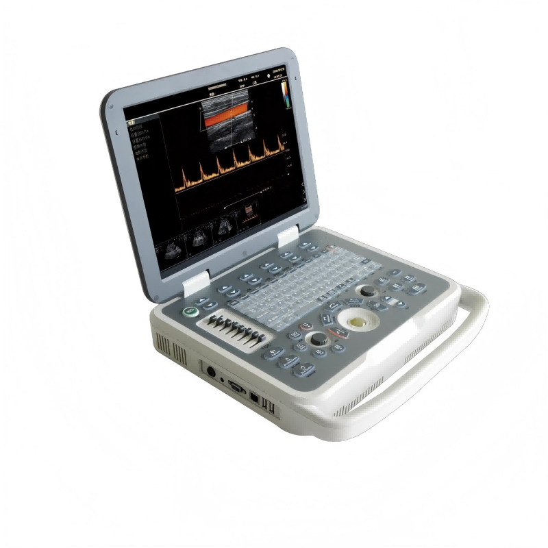
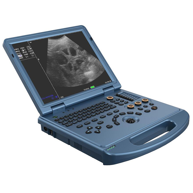
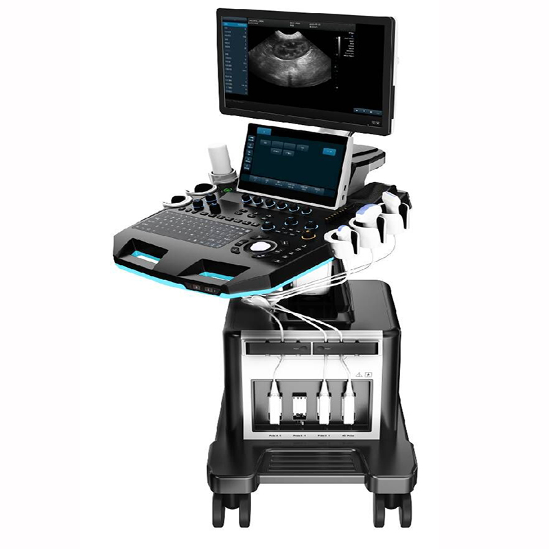
Related products
See all →
Portable Veterinary Color Doppler Ultrasound Scanner VC-E80
VC-E80 Portable Veterinary Color Doppler Ultrasound Scanner
Trolley Veterinary Color Doppler Ultrasound Scanner VC-U9
VC-U9 Trolley Veterinary Color Doppler Ultrasound Scanner
Portable Veterinary Color Doppler Ultrasound Scanner VC-U60
VC-U60 Portable Veterinary Color Doppler Ultrasound Scanner

Portable Color Doppler Ultrasound Scanner VC-U6
VC-U6 Portable Color Doppler Ultrasound Scanner
Phased Array Probe Type Wireless Color Doppler Scanner VC-4PA
VC-4PA Phased Array Probe Type Wireless Color Doppler Scanner
Micro-Convex+Linear Probe Type Wireless Color Doppler Scanner VC-3ML
VC-3ML Micro-convex+Linear Probe Type Wireless Color Doppler Scanner
Micro-convex Probe Type Wireless Color Doppler Scanner VC-3M
VC-3M Micro-convex Probe Type Wireless Color Doppler Scanner